
Peripheral arteriovenous malformations: questions and answers
Peripheral vascular malformations are congenital abnormalities of the blood vessels in the limbs and soft tissue, treatable percutaneously without surgery. Here are the answers to the most common questions about diagnosis and treatment — a different topic from cerebral arteriovenous malformations, which are managed by neurosurgery.
Do you recognise these symptoms?
- Visible or palpable swelling, present since birth or childhood
- Pain or a sense of tension in the affected area
- Skin patches associated with the malformation
- Limited movement if the malformation involves muscles or joints
- Tingling or altered sensation if a nearby nerve is compressed
What are peripheral vascular malformations?
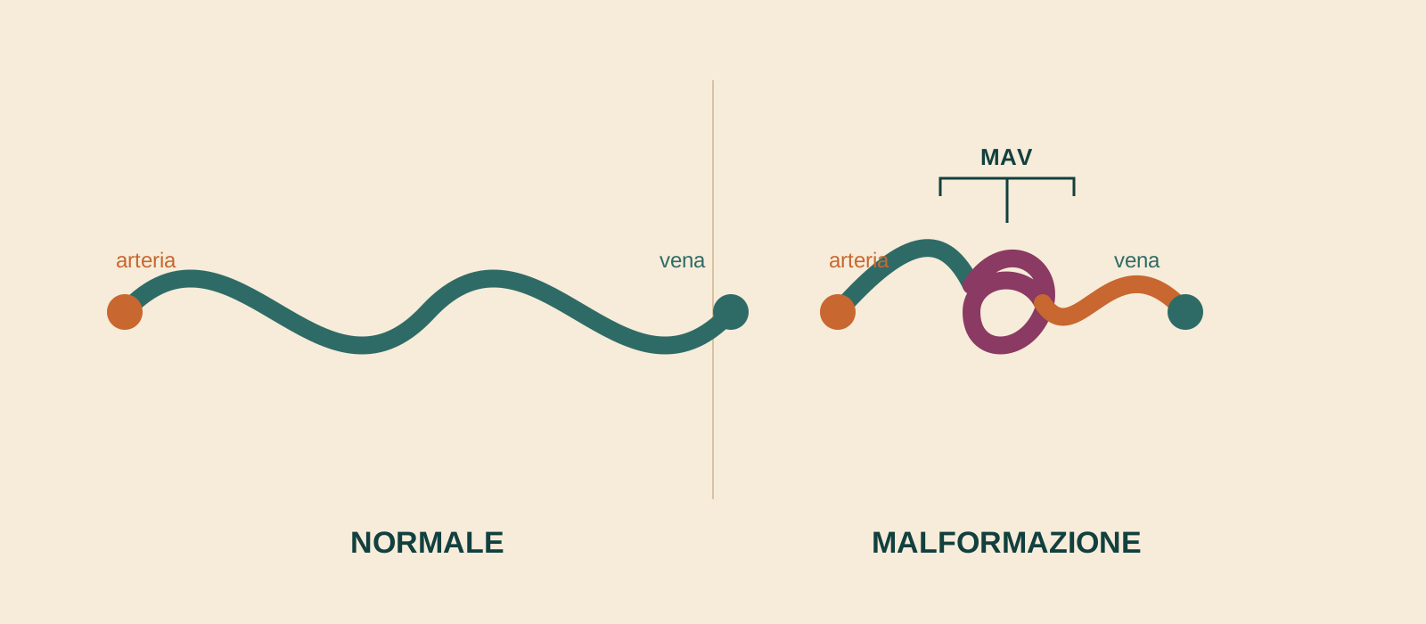
They are congenital abnormalities in the formation of blood vessels, present from birth even though they sometimes only become evident later. They fall into two broad groups: low-flow malformations (venous, lymphatic or veno-lymphatic, involving only veins or lymphatic vessels) and high-flow malformations (arteriovenous, in which arteries and veins communicate directly without the normal capillary network).
How are they diagnosed?
The first test is a Doppler ultrasound, which allows the type of malformation to be suspected and distinguishes low-flow from high-flow forms. MR angiography or CT angiography are used to better characterise the extent; arteriography remains the specific test for studying arteriovenous malformations in detail and planning treatment.
How are they treated percutaneously?
Through a simple puncture, a thin catheter is guided using radiological imaging to the malformed vessels; embolic or sclerosing agents are then injected to block the abnormal flow, reducing the malformation. No surgical incision is required.
What to expect from treatment?
An overnight hospital stay is usually required. Some discomfort in the treated area is normal in the three days following the procedure. Because malformations can be extensive or complex, treatment often needs to be repeated several times, about six weeks apart, until the desired result is achieved.
Frequently asked questions
What is the difference between low-flow and high-flow malformations?
Low-flow forms (venous, lymphatic) involve only veins or lymphatic vessels; high-flow forms (arteriovenous) directly connect arteries and veins, bypassing the normal capillary network, and generally require a different treatment approach.
Are vascular malformations always painful?
No: many remain asymptomatic for years. Others cause swelling, pain or functional limitations depending on their location and extent.
What does treatment involve?
A thin catheter is guided using radiological imaging, through a simple puncture, to the malformed vessel: agents that block the abnormal flow are then injected. No surgical incision is needed.
Is hospital admission needed?
Usually yes, for one night of observation after the procedure.
Is one session enough?
Not always: for more extensive forms, treatment may need to be repeated several times, about six weeks apart.
Does it hurt after the procedure?
Some discomfort in the treated area is normal in the three days following it.
Is this the same as cerebral arteriovenous malformations?
No: this covers peripheral vascular malformations of the limbs and soft tissue, treated by interventional radiology. Cerebral arteriovenous malformations are a different condition, managed by neurosurgery and neuroradiology.
Want to know if it is your case?
At the first consultation we review your clinical history and previous imaging together.
Other guides
The information on this page is for general guidance and does not replace a medical consultation. The indication for treatment must always be assessed individually.